Artificial Intelligence is now used in a variety of industries, including healthcare. In the field of healthcare, it has been used to perform tasks such as testing drug interactions or estimating treatment effects using prediction and probability, extracting information from unstructured medical text using natural language processing (NLP), diabetic retinopathy, and XRay/CT scan analysis using image processing and deep learning, risk assessment, and so on.
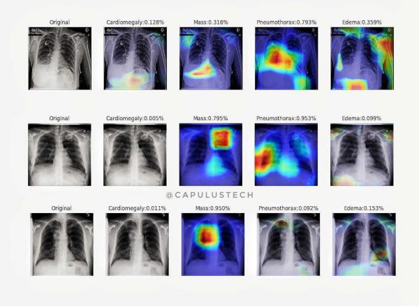
Chest X-rays are the most popular imaging test method used in medicine, with approximately 2 billion procedures each year. They are important for the screening, diagnosis, and treatment of diseases such as pneumonia. But, two-thirds of the world’s population is believed to be without access to radiology diagnostics.
The hope is that by automating this process at the expert level, this technology can enhance healthcare delivery and expand access to medical imaging knowledge in areas where radiologists are very limited. Here comes the deep learning method to analyze the medical image and give results with radiologist performance level (source).
COVID-19, also known as Novel Coronavirus Disease, is a highly infectious disease caused by SARS-CoV-2 that belongs to the broad family of coronaviruses. The disease first appeared in Wuhan, China in December 2019 and quickly spread to over 213 plus countries, becoming a global pandemic. Fever, dry cough, and exhaustion are the most common COVID-19 symptoms. Aches, pains, and trouble breathing are some of the other symptoms that people can experience. The majority of these symptoms are signs of respiratory infections and lung defects, which radiologists can identify.
So far deep learning has been used to detect several lung diseases using X-ray/CT scans. In particular, detection of Lung Cancer, Pneumothorax, Edema, Mass, etc(have a look).
Since covid-19 is also affecting the lungs, an X-ray or CT scan of the lungs can be used to predict whether the person is covid positive or not. Making it a faster way to give the test results. The dataset which is used to implement this is from opensource repositories (chest Xrays, chest ct-scans). This can be achieved by training the deep learning model over the x-ray datasets.
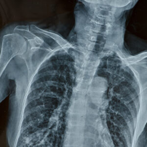
The next step is to Train the model, But before that, the dataset should be preprocessed. This is needed because the dataset is small and contains only a thousand images. The augmentation of these images provides a great advantage over the training of Artificial Nural Network(source). Once the images are split as train, test dataset, and augmented, the next is to choose the model to train. Here we have used a pre-trained model Densnet and transfer learning it with modifying its final layers by adding a global average pool and a dense layer of two neurons to predict which class(covid or no-covid) the Xray belongs to. The model is trained until it reaches 97% accuracy on the chest X-ray data. The same steps are repeated for the ct-scan image dataset. and the model is evaluated as follows.
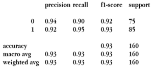
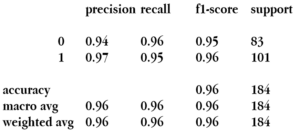
Model Classification report for CT-Scan(left) and Chest-Xray(right)
Model ROC Curve for CT-Scan(left) and Chest-Xray(right)
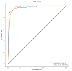
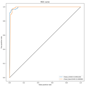
m the Receiver Operating Characteristics(ROC) curve and Classification Report of the model, it is clear that the model performing very well over the test data with an average accuracy of 95-96%. Here are the model results on x-ray and ct-scan images.
Image classification results for CT-Scan

Image classification results for Chest-Xray

